Description
The RGNeurov1 promoter is a compact, synthetic regulatory element designed for neuron-specific gene expression. At approximately 200 bp in length, this minimal promoter selectively drives neuronal expression while minimizing non-specific background. RGNeurov1 shows strong activity in neuronal contexts both in vitro and in vivo, with compatibility for AAV-based applications.
Key Features:
-
Tissue-Specific Expression – RGNeurov1 is designed to preferentially drive expression in neurons, modeled after Synapsin1 promoter activity.
-
Gene(s) of Interest – Validated using ZsGreen-myc reporter constructs
-
Validated Performance –
- Cell Culture: Expression observed in HEK293 and N2a cells; stronger neuronal expression in N2a
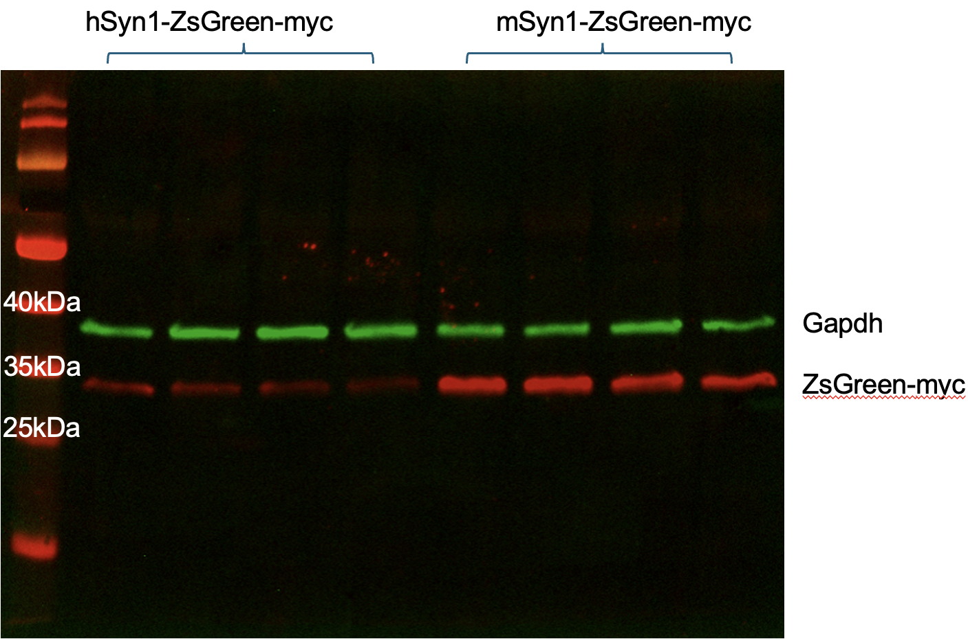
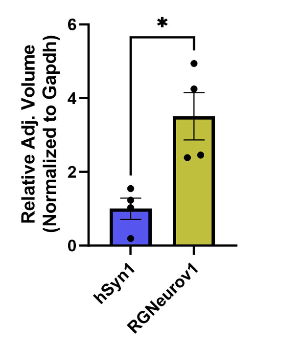
- Western Blot: Myc-tag detection confirms transgene expression; densitometry analysis shows robust promoter activity relative to hSyn1.
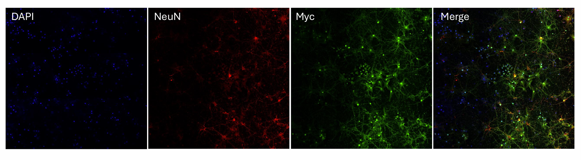
- Primary Neuron Culture: In mixed mouse cortical cultures, RGNeurov1 Selectively expressed neurons, where the ubiquitous CAG promoter did not.
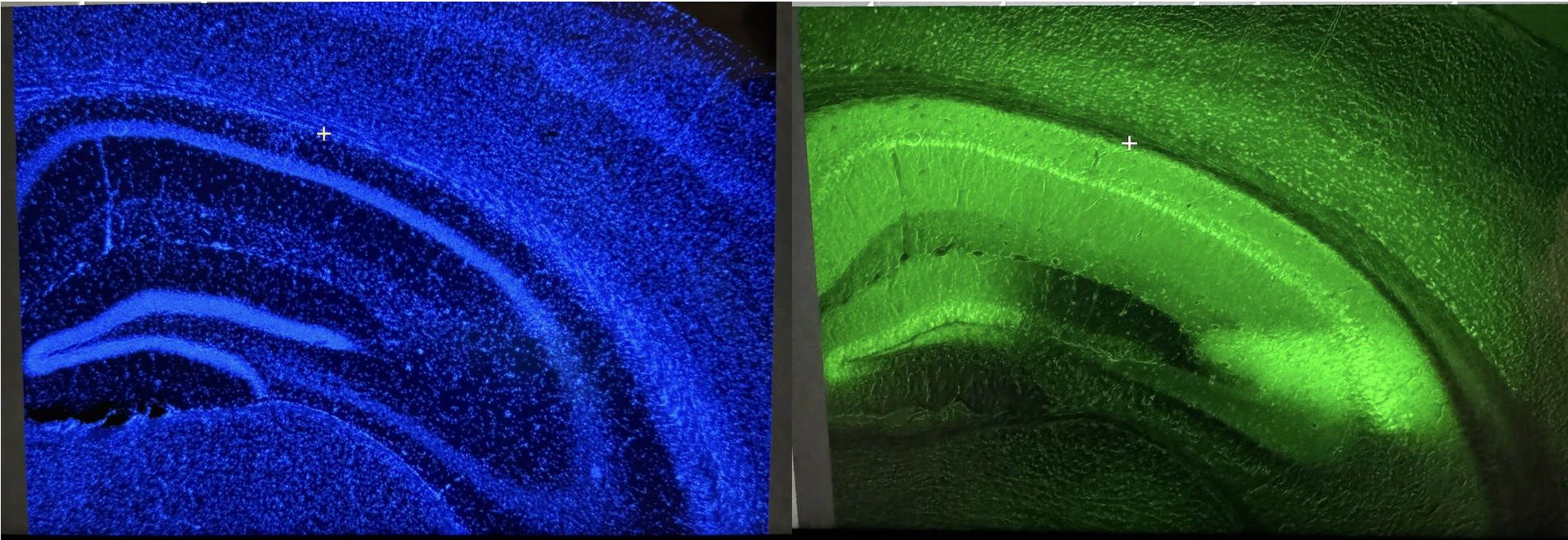
- In vivo: ssAAV9 delivery into adult mouse hippocampus yielded strong neuronal ZsGreen expressions
-
Compatibility – Tested in plasmid-based using Lipofectamine 3000 transfection and viral AAV9 systems, showing reliable performance across both
- References (Links to external research involving hsyn1):
- Widespread neuron-specific transgene expression in brain and spinal cord following synapsin promoter-driven AAV9 neonatal intracerebroventricular injection
- Human synapsin 1 gene promoter confers highly neuron-specific long-term transgene expression from an adenoviral vector in the adult rat brain depending on the transduced area
- Better Targeting, Better Efficiency for Wide-Scale Neuronal Transduction with the Synapsin Promoter and AAV-PHP.BUpdated
Validation Data: (Figures 1-11 are laid out in order from left to right)
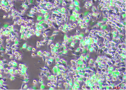
Figure 1: HEK293 cells were transfected with 500 ng of predicted RGNeurov1-ZsGreen-myc plasmid using Lipofectamine 3000 and imaged after 24 h incubation (fluorescence overlaid with brightfield)
Figure 2: HEK293 cells were transfected with 500 ng of hSyn1-ZsGreen-myc plasmid (VectorBuilder) using Lipofectamine 3000 and imaged after 24 h incubation (fluorescence overlaid with brightfield)
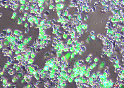
Figure 3: N2A cells were transfected with 500 ng of predicted RGNeurov1-ZsGreen-myc plasmid using Lipofectamine 3000 and imaged after 24 h incubation (fluorescence overlaid with brightfield)
Figure 4: N2A cells were transfected with 500 ng of hSyn1-ZsGreen-myc plasmid (VectorBuilder) using Lipofectamine 3000 and imaged after 24 h incubation (fluorescence overlaid with brightfield)
Figure 5: Western blot of HEK293 cell lysates transfected with hSyn1-ZsGreen-myc or RGNeurov1-ZsGreen-myc. GAPDH was used as a loading control, and myc staining was used to assess relative transgene expression and compare promoter strength.
Figure 6: Densitometry analysis showing fold change of ZsGreen-myc expression normalized to GAPDH. RGNeurov1 promoter drove significantly higher expression than hSyn1
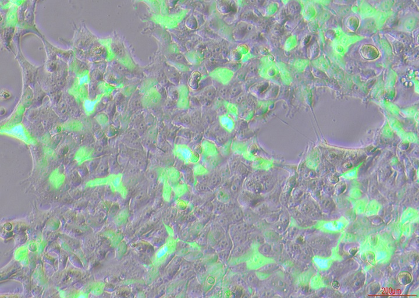
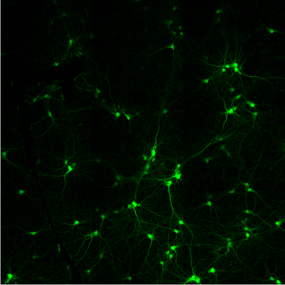
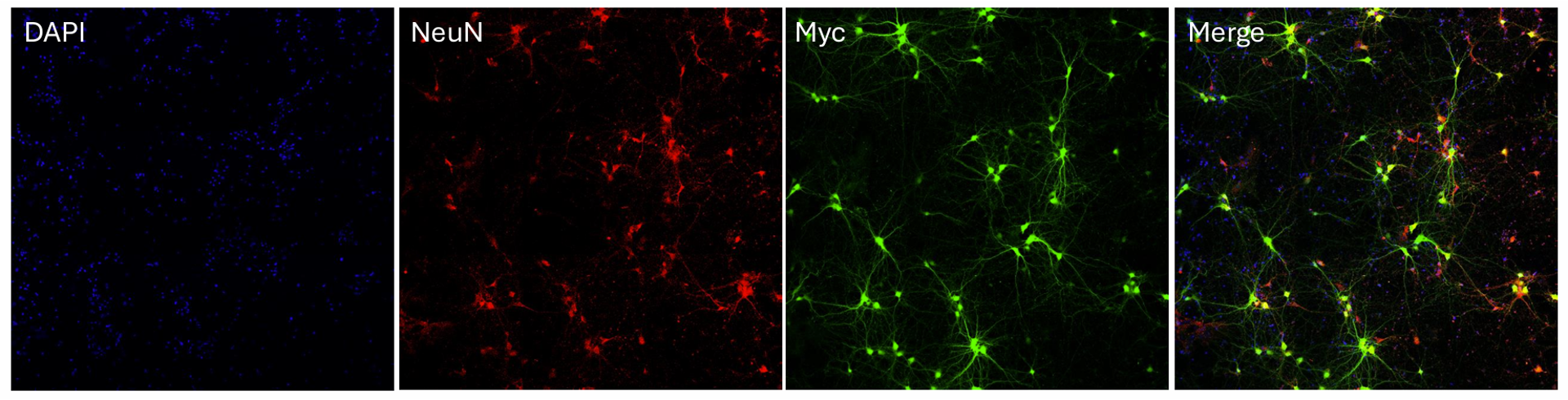
Figure 7: Neuron-specific expression from the RGNeurov1 promoter in primary cortical co-cultures transduced with ssAAV9-RGNeurov1-ZsGreen-myc (2000 vg)
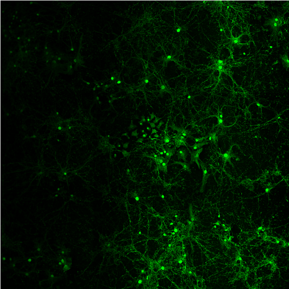
Figure 8: Broad expression from the CAG promoter in cortical co-cultures transduced with ssAAV9-CAG-GFP (2000 vg)
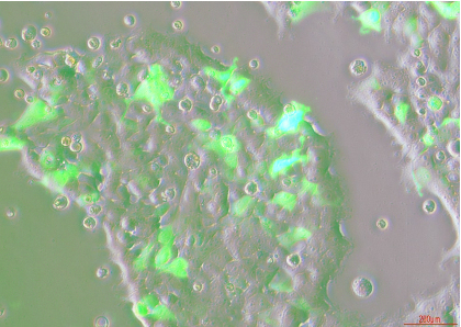
Figure 9: Broad expression with CAG promoter (25x magnification)
Figure 10: Neuron-specific expression with RGNeurov1 promoter (25x magnification)
Figure 11:
- Left image (DAPI): DAPI expressions in the hippocampus of the adult male mouse brain treated with ssAAV9-RGNeurov1-ZsGreen-myc via intra-hippocampal injections (10x magnification).
- Right image (ZsGreen): ZsGreen signal in the hippocampus of the adult male mouse brain treated with ssAAV9-RGNeurov1-ZsGreen-myc via intra-hippocampal injections (10x magnification).
Credits: Validation data provided by Re:Pair Genomics
Applications:
-
Neuroscience Research
-
Functional Genomics
-
AAV-based Gene Delivery
-
Translational Neuroscience